Circulatory system of the lancelet: features of the structure
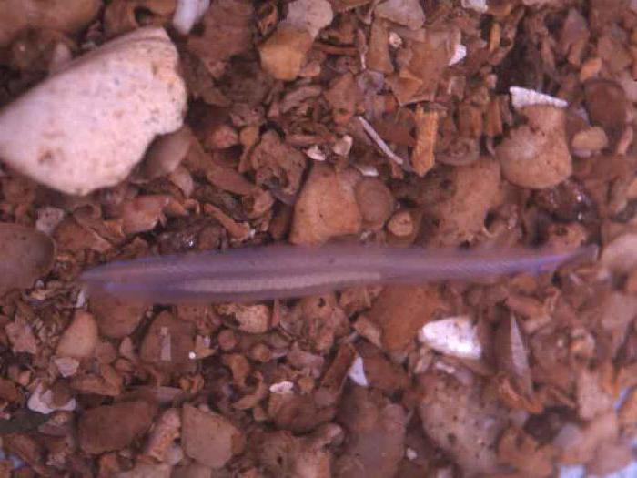
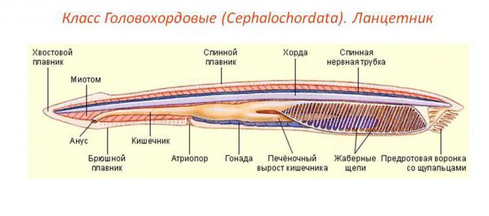
On the sandy bottom of the seas lead a bottom-line lifewhite-cream or slightly pinkish translucent animals, called lancelets. Their sizes are from 5 to 8 cm. The body is flattened from the sides, the front end of it is obliquely cut off, and on it is a mouth framed by tentacles. The back of the body looks like a surgical knife - a lancet. Such comparatively anatomical animals and zoology are studied quite seriously for one reason: the lancelet is considered to be the connecting link between two important groups of animals, invertebrates and chordates.
In this paper, we compare the structure of the lancet withbone fishes, and also give an answer to the following question: which circulatory system in the lancelet? Russian biologist AO Kovalevsky in 1860 proved that this animal has features similar to vertebrates, preserving the signs of invertebrate organisms.
Circulation Circle
Consider the structure of the circulatory systemlancelet. The red liquid, which does not have pigments, moves along the abdominal aorta, which constantly pulsates due to contractions of the myoepithelial layer of the cavity of the coelom. Then the blood with an excess of carbon dioxide enters the head of the lancelet. Gas exchange takes place in the gill vessels. Arteries flow into the posterior pharynx, where the right and left sections of the dorsal aorta are located. The anterior part of the body of the lancelet is provided by blood from the carotid arteries emerging from the aorta. For smaller arterioles, oxygen-rich blood enters all organs of the animal. The venous part of this system begins with a network of intestinal venules containing carbon dioxide. Of these, blood enters the swine vein.

Here, the portal system of the liver is formed. Anatomically, it is located under the intestinal tube of the lancelet, decaying into a network of venules that braid the walls of the digestive system. Its function is to transfer the toxified blood with a high content of carbon dioxide further into the venous sinus. From both parts of the body of the lancelet, it goes to the cardinal (otherwise called jugular) veins, then into the Cuvierian ducts.
Cuvierian ducts
These veins of vertebrates for the first timeIsolate at the lancelet and are formed by the fusion of cardinal vessels. In them, the red liquid comes from the front and back ends of the animal's body. Cuvier ducts directly flow into the venous sinus, which is considered the beginning of the abdominal aorta. These vessels are clearly expressed in embryos of vertebrates, and in the postembryonic period they are characteristic of cyclostomes (lampreys and myxins), as well as fish and amphibians. The major features of similarity are the circulatory system of lancelet and cyclostomes, although the latter have a real heart, consisting of the atrium and the ventricle.
Venous sinus
It is the initial part of the abdominal aorta, andsuch a lancelet system is a vicious circle. Thus, the structure of the lancete circulatory system proves that its blood circulation is closed. In mammals, birds and other vertebrates this part of the organs belongs to the right atrium. From it, the venous fluid enters the ventricle and then into the pulmonary arteries. So begins a small circle of blood circulation in organisms that have a four-chambered heart. In the lancelet, like the other representatives of the cephalothorax, the heart is absent and the venous sinus is represented by an unpaired vessel into which the venous fluid comes from the hepatic vein. Then it passes into the abdominal aorta. If you recall the structure of the blood system of the lancelet and bony fishes, you will find that the changes have primarily affected the abdominal aorta, which in fish is modified into a two-chambered heart. In addition, the respiratory surface of the gills of bone fish also increased due to the branching of the capillary network of their gill arteries.

Gate system of hepatic outgrowth
The circulatory system of the lancelet, like othersvertebrate animals, is anatomically linked to the digestive organs. Digestive all vertebrate animals are morphologically related, and the products of dissimilation: glucose, amino acids - come into its capillaries. Continuing to study the structure of the circulatory system of Amphioxus, adding that all of the liquid from the animal's digestive apparatus enters the hepatic outgrowth. Similarly, the liver of fish, amphibians and other vertebrates, Amphioxus this body performs the function of detoxification, cleansing the blood coming from the intestines from the decay products - metabolites. Then it enters the venous sinus. We add that the hepatic outgrowth of the blood comes from the sub-intestinal vein.
Abdominal and dorsal aorta
It is the main arterial vessel. If you remember the structure of the lancet circulatory system, then on the micropreparation you will see that under the throat of the animal there is the abdominal aorta, from which the pair arteries symmetrically depart. They branch into the septa of the gill cavities. The dorsal aorta forms at the posterior end of the pharynx as a result of the fusion of the epicarbral arteries. Anatomically, it is located under the chord and extends to the posterior end of the body of the lancelet, branched into arteries feeding the internal organs of the animal. In the lancelet, the metabolic products in the blood are filtered using special tubes called protenephridia. From the abdominal aorta to the body cavity - the whole - the arterial vessel is suitable. It branches into capillary glomeruli. Through their walls, plasma filtration takes place, and toxins are dissolved in dissolved form in protenephridia, then into the meso-neural duct and then into the cloaca.
The circulatory system of the lancelet and bony fishes
Consider the features of similarity and differences in structurecardiovascular system of the supra-class Bone fishes and the type of the head-headed, to which the lancelet belongs. Both groups of animals have one circle of blood circulation. But the lancelet does not have a heart, its function takes over a part of the abdominal aorta, which contracts together with the gill arteries and creates a flow of blood. The fish have a heart, it, like the cyclostomes, has a two-chamber (atrium and ventricle).

The formation of this body is associated with a more activemetabolism. The heart of the fish is located next to the isthmus under the lower jaw. As we have seen from the above facts, the structure of the circulatory system of the lancelet, which provides the transfer of oxygen and nutrients, differs from that of bone fish.
Features of the blood supply of the gill apparatus
If you remember the structure of the circulatory systemlancelet, compare it with bone fishes, then you will find differences that have affected the blood supply of the gill apparatus. The abdominal aorta is located on the lower side of the pharynx. From it to each pair of branchial arches arteries approach, which carry venous blood. The decrease in the number of septa in the gills (150 pairs in the lancelet and 4 pairs in the fish) is explained by increased metabolism, as well as an increase in the total area of the capillary network in representatives of bone fishes. The lancet is able to saturate its blood with oxygen, not only through the system of the gill arteries, but also by direct diffusion of gas through the skin into the superficial blood vessels.

Sleepy arteries
If you compare the blood systems of the lancelet andbone fish, then you will find differences regarding the vessels called the carotid arteries. They carry the arterial red fluid to the anterior end of the animal body. In bone fish, 4 pairs of gill arteries flow into the dorsal aorta, the roots of which separate the carotid arteries. In the lancelet, the number of gill vessels is much larger. They supply oxygen to the brain, representing the expansion of the neural tube and not differentiated into departments. He controls the reflex activity of the animal. The provision of brain neurons with oxygen and nutrients is due to the branching of the carotid arteries to the capillary system. It also receives products - metabolites, sent through the veins into the venous sinus.
In this article, the circulatory system of the lancelet and the circulatory features of the cephalothorax have been studied.